Plantar Foot Muscles Mri / The muscles lying within the medial group form a bulge referred to as the 'ball' of the big toe.
byAdmin-
0
Plantar Foot Muscles Mri / The muscles lying within the medial group form a bulge referred to as the 'ball' of the big toe.. / muscles that move the foot and toes. The flexor digitorum brevis muscle lies superficially under the plantar aponeurosis and marks the largest muscle in the central compartment. 23,25 mri at the level of the malleolus demonstrates the muscle as. Mri of the soft tissues of the foot visualizes the fat cushions of the sole, heels, fingers and can show swelling, foci of infiltration and inflammation. The origins of the lumbrical muscles are located at the distal end of the quadratus plantae muscle.
The three plantar interossei muscles adduct the 3 rd, 4 th and 5 th toes toward the long axis through the 2 nd toe. Please come back soon to see the finished work! Imaging findings of tarsal tunnel syndrome depend on underlying etiology. Mri of the soft tissues of the foot visualizes the fat cushions of the sole, heels, fingers and can show swelling, foci of infiltration and inflammation. Nodules or masses of plantar fibromatosis are typically located in the middle to the medial aspect of the plantar arch and may extend to involve the skin or deep structures of the foot.
Mri Appearance Of Jogger S Foot Springerlink from media.springernature.com The plantar fascia is a multilayered, fibrous aponeurosis with medial, central, and lateral components (, 1).the term plantar fascia typically refers to the large central component, which originates from the medial calcaneal tuberosity and extends anteriorly, adhering to the underlying flexor digitorum brevis (fdb) muscle. It is considered the most common cause of heel pain. Medial sides of metatarsals of toes iii to v insertion: These patients usually present with heel or arch pain. It contributes to the surface anatomy of the medial sole of the foot and is easy to palpate. 23,25 mri at the level of the malleolus demonstrates the muscle as. Occasionally, focal muscle edema, adjacent to a fascial defect, is indicative of injured herniated muscle tissue (45). The transverse (adt) and oblique (ado) heads of the adductor hallucis muscle send fibers to the lateral sesamoid, capsule and plantar plate.
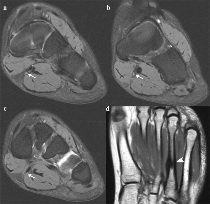
The flexor hallucis longus tendon (fhl) is also depicted.
/ muscles that move the foot and toes. 10 foot and ankle craig r. Smaller terminal division of the tibial nerve course: At about the midsole, it splits into five. The studies were performed on a variety of magnets ranging from 0.2 to 1.5 t between march 15 and july 22, 2006. It attaches to the lateral base of the proximal phalanx of the 5th digit. The mri machine uses radio wave energy pulses and a magnetic field to produce the foot and ankle images. 23 it can originate as a separate muscle from the fibula or from the peroneus brevis or longus muscles and inserts onto the peroneal tubercle or retrotrochlear eminence of the calcaneus. The lateral plantar nerve is also commonly entrapped at the tarsal tunnel or distal to the tarsal tunnel with loss of sensation along the distal third of the foot. The interosseous muscles of the fourth interspace are usually supplied by a branch from the superficial ramus of the lateral plantar interosseous, plantar the plantar interosseous muscle arises from the proximal third of the medial plantar surface of the shaft, from the base of the metatarsal on which it lies, and from the fascial expansions of. Imaging findings of tarsal tunnel syndrome depend on underlying etiology. Lesions may be symptomatic because of a mass effect or invasion of adjacent muscles or neurovascular structures. Abdm, abductor digiti minimi muscle;
Muscles that move the foot and toes. These patients usually present with heel or arch pain. The muscles lying within the medial group form a bulge referred to as the 'ball' of the big toe. In addition, an image of all the muscles of the back and plantar part of the foot, all tendons and tendon ligaments, blood vessels and nerves are obtained. Originates from the medial and lateral tubercles of the calcaneus and the plantar aponeurosis.
Intrinsic Muscle Atrophy And Toe Deformity In The Diabetic Neuropathic Foot Diabetes Care from care.diabetesjournals.org The flexor digitorum brevis muscle lies superficially under the plantar aponeurosis and marks the largest muscle in the central compartment. It is considered the most common cause of heel pain. The interosseous muscles of the fourth interspace are usually supplied by a branch from the superficial ramus of the lateral plantar interosseous, plantar the plantar interosseous muscle arises from the proximal third of the medial plantar surface of the shaft, from the base of the metatarsal on which it lies, and from the fascial expansions of. Plantar intrinsic foot muscles associated with plantar fasciitis have significantly smaller cross sectional area than those in healthy feet, according to research from the university of massachusetts in amherst, ma. The flexor hallucis longus tendon (fhl) is also depicted. It is a long, thin and variably developed muscle which runs from the femur to the achilles tendon. Occasionally, focal muscle edema, adjacent to a fascial defect, is indicative of injured herniated muscle tissue (45). The lateral plantar nerve is also commonly entrapped at the tarsal tunnel or distal to the tarsal tunnel with loss of sensation along the distal third of the foot.
The flexor digitorum brevis muscle lies superficially under the plantar aponeurosis and marks the largest muscle in the central compartment.
At mr imaging, the course of the plantar tendons is optimally visualized with dedicated imaging of the midfoot and forefoot. Nodules or masses of plantar fibromatosis are typically located in the middle to the medial aspect of the plantar arch and may extend to involve the skin or deep structures of the foot. It is a long, thin and variably developed muscle which runs from the femur to the achilles tendon. 23,25 mri at the level of the malleolus demonstrates the muscle as. Are plantar fasciitis and heels spurs the same thing? Head, neck, arm, foot, pelvis, etc. Chronic plantar fasciitis may be accompanied by muscle atrophy of plantar intrinsic foot muscles and tibialis posterior compromising the dynamic support of the foot prolonging the injury. In addition, an image of all the muscles of the back and plantar part of the foot, all tendons and tendon ligaments, blood vessels and nerves are obtained. Medial sides of metatarsals of toes iii to v insertion: The three groups of plantar foot muscles are(14): The three plantar interossei muscles adduct the 3 rd, 4 th and 5 th toes toward the long axis through the 2 nd toe. Familiarity with the normal anatomy of the plantar tendons and its appearance at magnetic resonance (mr) imaging and ultrasonography (us) is essential for recognizing plantar tendon disorders. Lesions may be symptomatic because of a mass effect or invasion of adjacent muscles or neurovascular structures.
Muscles that move the foot and toes. / muscles that move the foot and toes. The transverse (adt) and oblique (ado) heads of the adductor hallucis muscle send fibers to the lateral sesamoid, capsule and plantar plate. In the past, these bone spurs were often blamed for heel pain and removed surgically. Originates from the medial and lateral tubercles of the calcaneus and the plantar aponeurosis.
Baxter 039 S Neuropathy Isolated Fatty Atrophy Of The Abductor Digiti Minimi Muscle In Association With Plantar Fasciitis Eurorad from www.eurorad.org It is homologous with the abductor digiti minimi of the hand. Smaller terminal division of the tibial nerve course: The studies were performed on a variety of magnets ranging from 0.2 to 1.5 t between march 15 and july 22, 2006. Familiarity with the normal anatomy of the plantar tendons and its appearance at magnetic resonance (mr) imaging and ultrasonography (us) is essential for recognizing plantar tendon disorders. Magnetic resonance images of the foot may be digitized to quantify muscle architecture. In the past, these bone spurs were often blamed for heel pain and removed surgically. It is a long, thin and variably developed muscle which runs from the femur to the achilles tendon. Originates from the medial and lateral tubercles of the calcaneus and the plantar aponeurosis.
Lesions may be symptomatic because of a mass effect or invasion of adjacent muscles or neurovascular structures.
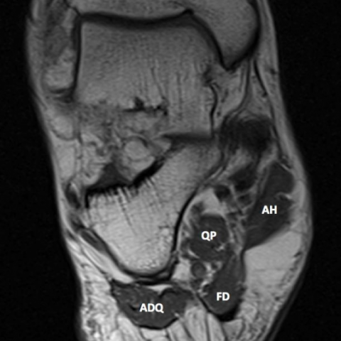
Plantar fasciitis refers to inflammation of the plantar fascia of the foot. The interosseous muscles of the fourth interspace are usually supplied by a branch from the superficial ramus of the lateral plantar interosseous, plantar the plantar interosseous muscle arises from the proximal third of the medial plantar surface of the shaft, from the base of the metatarsal on which it lies, and from the fascial expansions of. Smaller terminal division of the tibial nerve course: The abductor digiti minimi muscle is located on the lateral side of the foot. The flexor digitorum brevis muscle lies superficially under the plantar aponeurosis and marks the largest muscle in the central compartment. The lateral plantar nerve is an important motor nerve in the foot because it innervates all intrinsic muscles in the sole, except for the muscles supplied by the medial plantar nerve (abductor hallucis, flexor digitorum brevis, flexor hallucis brevis, and first lumbrical). The flexor hallucis longus tendon (fhl) is also depicted. In addition, an image of all the muscles of the back and plantar part of the foot, all tendons and tendon ligaments, blood vessels and nerves are obtained. Medial sides of metatarsals of toes iii to v insertion: Certain soft tissue tumours are identifiably benign because of their signal characteristics, morphology and/or location. Familiarity with the normal anatomy of the plantar tendons and its appearance at magnetic resonance (mr) imaging and ultrasonography (us) is essential for recognizing plantar tendon disorders. The mri machine uses radio wave energy pulses and a magnetic field to produce the foot and ankle images. The peroneus quartus muscle is more common, presenting in 13% to 22% of the population.
It is a long, thin and variably developed muscle which runs from the femur to the achilles tendon foot muscles mri. The plantar fascia is a multilayered, fibrous aponeurosis with medial, central, and lateral components (, 1).the term plantar fascia typically refers to the large central component, which originates from the medial calcaneal tuberosity and extends anteriorly, adhering to the underlying flexor digitorum brevis (fdb) muscle.